Peter M. Clark, Ph.D.
- Associate Professor, Molecular and Medical Pharmacology
- Associate Professor, UCLA Crump Institute for Molecular Imaging

Peter M. Clark, Ph.D., aims to develop new therapies for cancer and autoimmune diseases. His research leverages molecular imaging technologies to discover critical pathways in these diseases and high-throughput screening platforms to identify new ways to target these pathways.
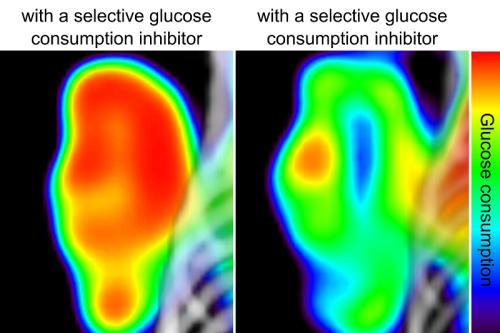
Applying his training in mathematics, biology and chemistry, Clark seeks to develop, validate and use new assays to improve the diagnosis and treatment of cancer. He developed a high-throughput assay for measuring glucose consumption that is over 100-fold faster than traditional assays. This tool can evaluate thousands of small molecules or proteins at a time to uncover the mechanisms driving glucose consumption in cancer cells, informing the development of selective inhibitors that regulate this consumption.
Clark has also identified the metabolic enzyme deoxycytidine kinase, dCK, as a key and targetable enzyme required for aberrant T and B cell activation in models of autoimmune diseases, such as multiple sclerosis, as well as rarer conditions, such as acute disseminated encephalomyelitis, or ADEM. This work contributed to the novel dCK inhibitor TRE-515, earning FDA Orphan Drug Status for the treatment of ADEM, a rare and sometimes fatal neurologic disease that primarily affects children and occurs after a viral or bacterial infection.
Research Projects
- Developing PET tracers that can determine the fate of stem cell-derived liver cells, which could advance cell therapies for liver disease and replace whole organ donation
- Identifying and studying selective inhibitors of cancer cell glucose consumption
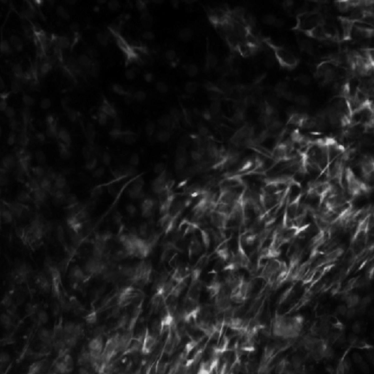
- Developing therapies for multiple sclerosis and other autoimmune conditions that work by depriving malfunctioning immune cells of a critical metabolic enzyme
-
Post-doctoral Fellowship
- Molecular Imaging, UCLA, 2015
Degree
- Ph.D., Chemical Biology, California Institute of Technology, 2010
-

