UCLA scientists find when and where human stem cells model the brain
Led by Dr. Daniel Geschwind, director of the UCLA Center for Autism Research and Treatment, scientists from UCLA’s Eli and Edythe Broad Center of Regenerative Medicine and Stem Cell Research have pioneered a set of biological data analysis (bioinformatics) tools that for the first time measure how successfully brain (neural) stem cells grown in the laboratory model the outermost part of the brain called the cortex during early stages of human fetal development. The nearly three-year study was published July 2, 2014 in the leading journal Neuron.
Drs. Luis de la Torre-Ubieta and Jason Stein, postdoctoral fellows in the lab of Dr. Geschwind and co-lead authors of the study, developed a comprehensive system that assesses neural stem cell differentiation across key data points including temporal identity (age range) and region of the brain. Stem cells are procured and incubated over a twelve-week period, and genes known to be representative of the cortex or that signify the transition of a stem cell into a neuron (marker genes) are used to identify individual biological processes. At several points during the process, the scientists use data techniques to profile every gene of the human brain. With these techniques, they are able to profile all phases of stem cell differentiation and compare it to human cortical development (from four weeks after conception to eighty years old).
Dr. Geschwind and his colleagues specifically investigated how in vitro developmental models could effectively model brain developmental processes during early fetal stages, which is a crucial time in the pathology of autism. Results showed that expression of autism genes is highly preserved in vitro.
“These new techniques offer extraordinary promise in the study of autism, because we now have an unbiased and genome-wide view of how genes are used in the development of the disease, like a fingerprint,” said Dr. Geschwind. “Our goal is to develop new treatments for autism, and this discovery can provide the basis for improved high-efficiency screening methods and open up an enormous new realm of therapeutic possibilities that didn’t exist before.”
“This particular study will have a wide range of impact,” added Stein. “Whatever disorder you are trying to model in a dish, whether it is autism or schizophrenia or even Alzheimer’s disease, the bioinformatics tools we have developed will allow scientists to assess how well their system models the actual human brain. This kind of analysis has never been done before, in this way.”
Drs. Stein and de la Torre-Ubieta have also created a machine-learning approach called CoNTExT that identifies the developmental maturity and regional identity of in vitro models, and assesses their strengths and weaknesses across multiple categories. CoNTExT has been made available via a user-friendly website and is designed as a resource for researchers worldwide.
“Our hope is that the scientific community will be able to use this particular program to create the best protocols and refine their methods,” said Dr. de la Torre-Ubieta. “We now have a gold standard of what is normal human brain development in vivo, and this online resource provides access to a whole picture of how the brain develops from a transcriptomic point of view.”
Dr. de la Torre-Ubieta’s research on this project was supported by the California Institute of Regenerative Medicine (CIRM), the state's stem cell research agency, training grant. Additional funding was provided by the UCLA Broad Stem Cell Research Center Innovation Award through philanthropy and other sources.
Meet the Researchers
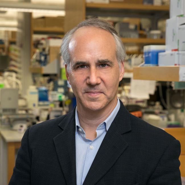
- Gordon and Virginia MacDonald Distinguished Professor, Human Genetics, Neurology and Psychiatry
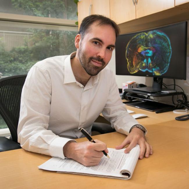
- Assistant Professor, Psychiatry and Biobehavioral Sciences