
Stem cell-derived brain models offer a new window into how anesthesia works
Key Takeaways
- UCLA researchers showed for the first time that lab-grown human brain assembloids can reproduce the electrical hallmarks of general anesthesia — the slow, sweeping brain waves that appear even as individual neurons fall quiet.
- By exposing the assembloids to the anesthetic propofol, the team found that a minimal circuit of human cortical neurons is enough to produce these changes, without the deeper brain structures long thought to be required.
- The work gives researchers a controllable human system for studying how anesthesia reshapes the brain's electrical rhythms — and potentially for investigating disorders that produce similar changes in brain activity.
Every day, hundreds of thousands of people undergo general anesthesia, trusting they'll drift into unconsciousness and wake safely after surgery. Yet despite decades of use, scientists still don't fully understand how anesthetic drugs reshape the brain's electrical activity to produce unconsciousness.
Studying the brain’s response to anesthesia has been difficult: in living humans, there’s no safe way to study the brain cell by cell, and in animals, the drugs act on many different brain regions at once, making it hard to isolate where the effects arise.
Now, UCLA researchers have shown for the first time that human stem cell-derived brain assembloids — tiny, three-dimensional models that recreate simplified human brain circuits — can reproduce the electrical changes seen during general anesthesia. Their findings were just published in the British Journal of Anaesthesia.
These new findings demonstrate that brain assembloids can serve as a human model for investigating how molecular changes inside brain cells shape large-scale patterns of electrical activity.
“We’re excited about these models’ potential not just for studying anesthesia, but as a tool we can use more broadly to understand how brain networks become disrupted in other disorders that profoundly alter brain dynamics like traumatic brain injury,” said senior author Dr. Ranmal Samarasinghe, an assistant professor of neurology and member of the UCLA Broad Stem Cell Research Center.
Scientists have long understood two pieces of the anesthesia puzzle: that drugs such as propofol bind to specific receptors on neurons, and what an anesthetized brain looks like on an electroencephalogram, or EEG.
“We’ve known what these drugs bind to for a long time, and we’ve known what happens at the level of the whole brain,” Dr. Daniel Toker, first author of the paper and a project scientist in Samarasinghe’s lab, said. “What we've been missing is a human model that lets us connect those two scales.”
Assembloids help close that gap. Unlike neurons grown flat in a lab dish, which are among the models that have previously been used in these types of studies, the assembloids contain interconnected circuits that produce coordinated electrical activity. And unlike animal brains, they let researchers isolate and manipulate those circuits in a controlled human system.
Slow waves, quiet neurons
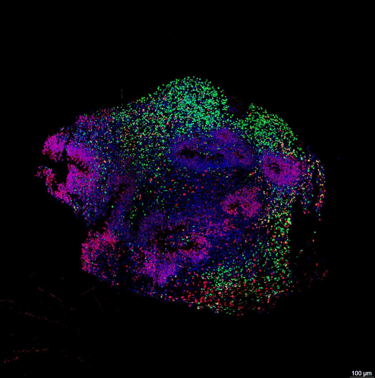
Created from induced pluripotent stem cells — adult cells that have been reprogrammed into a stem cell-like state — the assembloids combine three types of brain cells: excitatory neurons, which transmit signals; inhibitory neurons, which restrain activity; and glial cells, which play multiple roles in brain function. Together, these cells form circuits capable of generating coordinated rhythms similar to those in the human brain.
To test whether the models could capture the effects of anesthesia, the team exposed them to propofol. Because assembloids more closely resemble the developing fetal brain than the mature adult brain, it was unclear whether they would respond the same way.
But they did. Researchers found that the assembloids developed the broad, slow brain waves characteristic of anesthesia, even as the electrical activity of individual neurons grew markedly quieter.
Those two changes may sound contradictory, but together they capture one of anesthesia's defining features.
“It’s a little counterintuitive,” Toker said. “Individual neurons become less active, but the overall brain waves become larger, because many neurons begin changing their activity in synchrony.”
Toker compares the effect to a football stadium. If thousands of spectators are all holding separate conversations, a single microphone picks up little more than background noise. But if everyone falls quiet and then claps together every few seconds, those synchronized bursts become impossible to miss. In much the same way, anesthesia quiets individual neurons while synchronizing activity across the network, producing the large, slow waves seen on an EEG.
The team confirmed that the response depended on the biological mechanisms identified in decades of animal research. Blocking the receptors targeted by propofol eliminated the effect, and simpler models lacking inhibitory neurons failed to produce the characteristic waves.
The findings also speak to a longstanding debate over whether these signatures require input from the thalamus, a deep brain structure that relays signals to the cortex. Because assembloids contain no thalamus yet still reproduced the pattern, the results show that a minimal cortical circuit can be sufficient on its own.
A platform beyond anesthesia
Beyond advancing scientists’ understanding of anesthesia, the study also expands what researchers can investigate using brain assembloids.
Because assembloids are built from human cells and can be precisely manipulated in the laboratory, researchers can now investigate how specific genes, cell types and signaling pathways shape coordinated brain activity — questions that have been difficult or impossible to study directly in people.
The model could also help screen candidate drugs and examine why patients differ in their sensitivity to anesthesia, including the rare cases of unintentional awareness during surgery.
More broadly, by showing that a lab-grown human brain model can shift between distinct states of electrical activity, the study opens a new way to investigate how human brain circuits generate — and change — their activity.
“What makes brain assembloids so powerful is that you build the circuit from the ground up and decide what goes into it,” Toker said. “Because we're building it ourselves, we have a real chance to understand what shapes the brain activity we see in patients under anesthesia and across different conditions from coma to traumatic brain injury to epilepsy.”
Notes
This research was supported by the National Institutes of Health, CURE Epilepsy, the International SCN8A Alliance, the Simons Foundation, the UCLA Intellectual and Developmental Disabilities Research Center, a UCLA Broad Stem Cell Research Center Innovation Award, the In Memory of Christina Louise George Fund and the Michael R. Bloomberg Revocable Trust.
Contact

Meet the Researchers

- Assistant Professor-in-Residence, Neurology