
Announcing the winners of the UCLA Broad Stem Cell Research Center’s 2024 microscopy image and video contest
The Eli and Edythe Broad Center of Regenerative Medicine and Stem Cell Research at UCLA is thrilled to announce the winners of its 2024 light microscopy image and video contest.
Now in its third year, this competition celebrates the artistry and innovation of researchers across UCLA using microscopy to reveal the unseen wonders of biology. After careful evaluation by a panel of scientists and microscopy experts, the five winning entries were selected for exhibiting a combination of technical mastery, visual impact and scientific relevance. Here are this year’s winners:
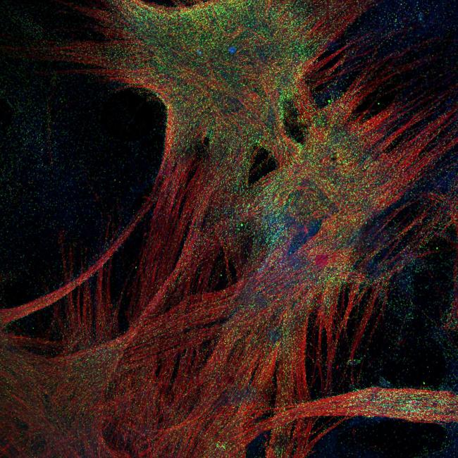
FIRST PLACE IMAGE
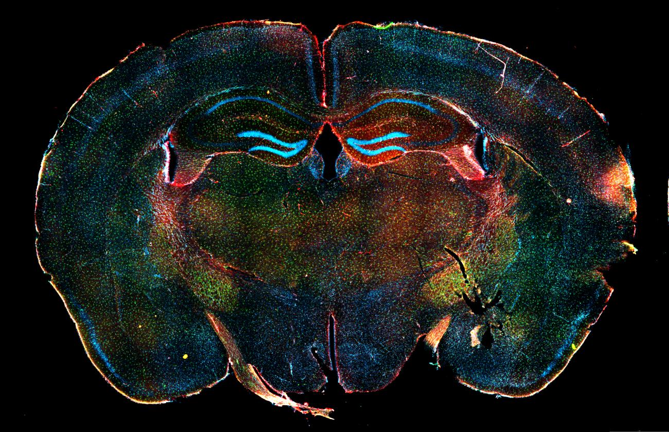
Title: Protectors of our brain
Instrument: Leica DMi8
Magnification: 10x
Creator: Rachel Reyes, Ph.D., a project scientist in the lab of Gerald Lipshutz, M.D., and Grace Wu
This is a microscopic image of a mouse brain displaying the main defensive players of our central nervous system. Astrocytes (red) and microglia (green) protect our resident neural cells, such as neurons (aqua), from chemical and mechanical damage, intruders and sometimes even from itself. The Lipshutz lab identifies changes in these populations as they relate to rare genetic neurodevelopmental disorders, especially those associated with liver metabolic disease. Having a better understanding of these rare disorders will inform the development and clinical application of much-needed gene and cell-based therapies to treat them.
SECOND PLACE IMAGE
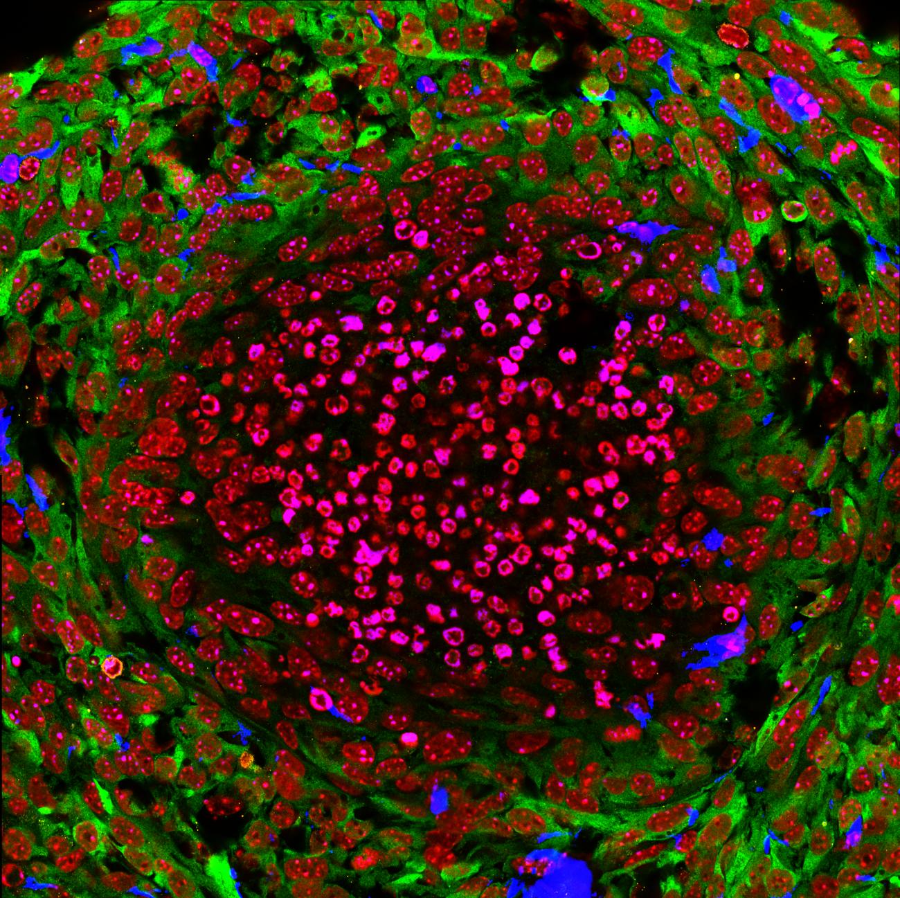
Title: Ring around the necrotic rosie
Instrument: Zeiss LSM 880
Magnification: 63x
Creator: Ryan Shih, a graduate student in the lab of Yvonne Chen, Ph.D.
The Chen lab seeks to develop cell therapies for difficult-to-treat solid tumors, such as glioblastoma. This requires that we understand why some therapies work and others fail. The image shown here is a cross-section of a brain tumor nodule in a mouse that did not respond to therapy. Cell nuclei (red) assume different morphologies in the dead core versus the surrounding living tumor shell (green). Different immune cells (blue and orange) are sparse, suggesting the therapy was ineffective in recruiting enough immune cells to mount an antitumor response. These results enable the lab to pinpoint how to optimize the cell therapies that they hope to one day bring to the clinic.
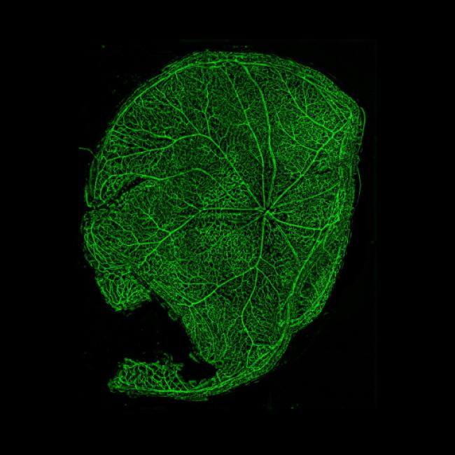
PEOPLE'S CHOICE IMAGE
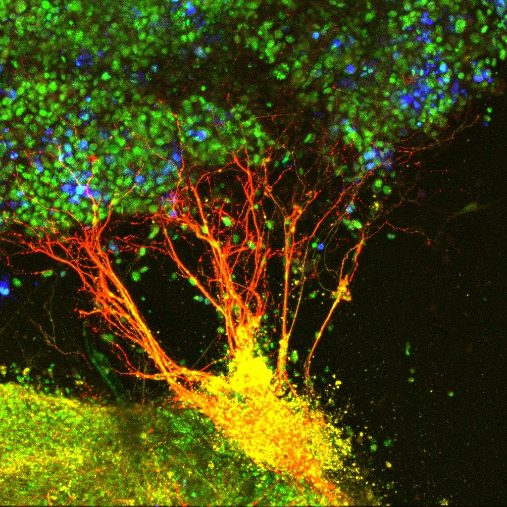
Title: A tree made of neurons
Instrument: Zeiss LSM 700
Magnification: 20x
Creator: Yesica Mercado-Ayon, a graduate student in the lab of Samantha Butler, Ph.D.
This image shows a bundle of dorsal interneurons from the rat embryonic spinal cord (red and yellow) growing towards an aggregate of cells expressing the green fluorescent protein (blue with nuclei labeled green). These neurons encode critical sensory modalities, including proprioception, nociception and touch, which are central to the human experience — allowing us to hold our bodies in space, react to pain and interact with our environment. The Butler lab aims to understand the mechanisms that establish neuronal networks during development and then co-opt these mechanisms to regenerate diseased or damaged spinal circuits.
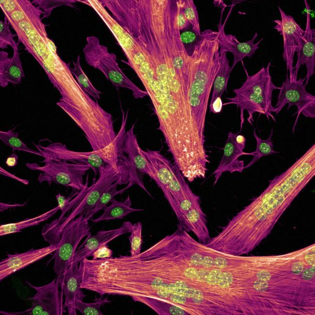
BEST UNDERGRADUATE SUBMISSION
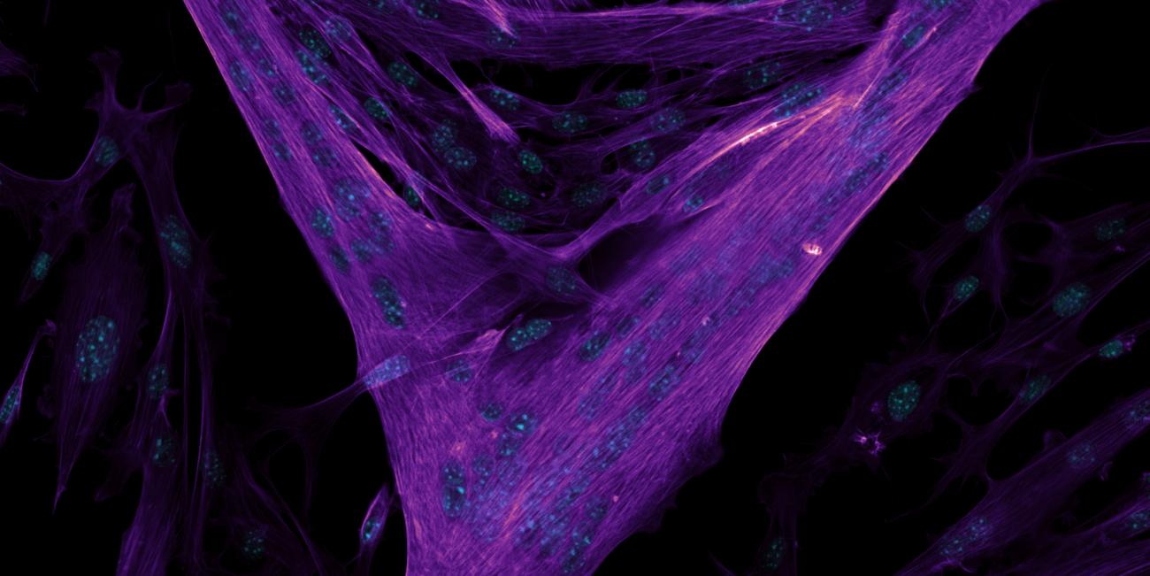
Title: Fiber frenzy
Instrument: Nikon AX R NSPARC
Magnification: 20x
Creator: Yutzil Herrera, a UCLA COMPASS trainee and an undergraduate student in the lab of Thomas Rando, M.D., Ph.D., and Tamas Nagy, Ph.D.
This microscopic image depicts muscle cells fusing to form myotubes with the actin cytoskeleton visible in purple and the nuclei in teal. Muscle fibers have the capacity to regenerate following injury, but as we age, these capabilities decline. These fusion assays are used in the Rando lab to study how muscle cells regenerate and how their interactions influence fusion. Studying these cellular processes contributes to our understanding of the biological changes observed in myogenic fusion as we age and will help the lab develop new therapies targeting conditions that lead to reduced muscle regeneration capacity.
FIRST PLACE VIDEO
Title: All aboard the (myo)train
Instrument: Cephla Squid
Magnification: 20x
Creator: Tamas Nagy, Ph.D., a postdoctoral scholar in the lab of Thomas Rando, M.D., Ph.D.
This video shows a muscle cell fusing into a proto-muscle fiber with the cell membranes visible in purple and yellow. Our muscles have a remarkable ability to regenerate after injury, but this ability declines as we age. Decades of research have uncovered many of the proteins and molecules involved in regeneration, but we still do not fully understand how these pieces come together to build healthy muscle. The Rando lab takes microscopic videos to understand how these molecular pieces influence the ways that muscle cells interact and coordinate the regeneration process. Gaining a better understanding of how this process breaks down as we grow older will enable scientists to design therapies to combat the effects of aging so that people live longer, healthier lives.
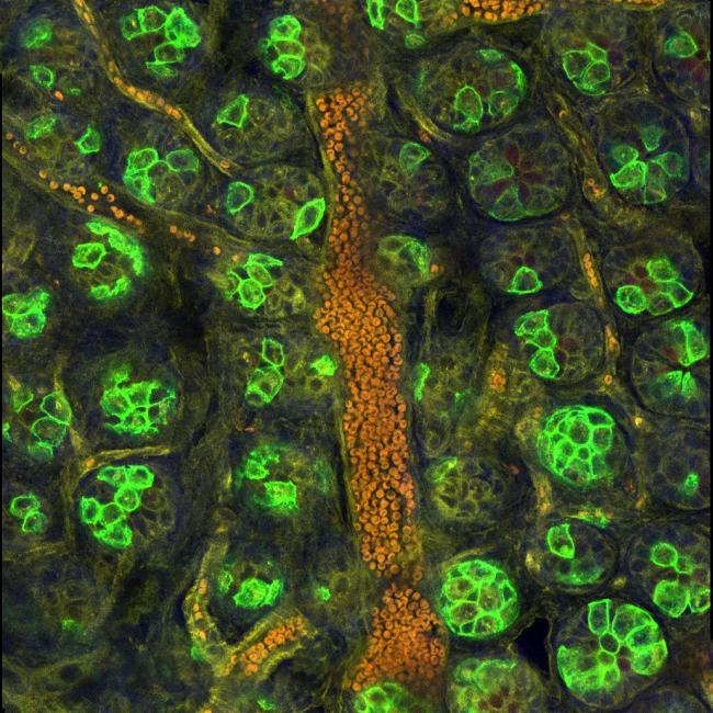
HONORABLE MENTIONS


Notes
The center would like to thank all who submitted images and videos as well as Nikon, who generously provided all the Nikon-branded prizes.
To see all contest submissions, visit the center's Flickr. The winning entries and the five images that received honorable mentions are listed first in the album.