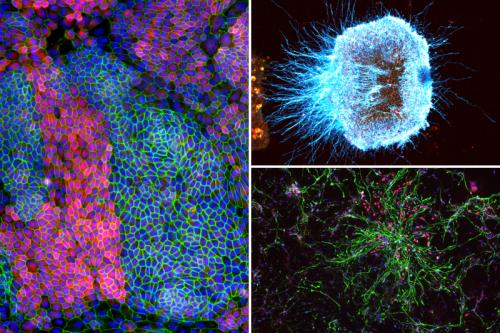
Enter the Broad Stem Cell Research Center's 2024 light microscopy image and video contest
The Eli and Edythe Broad Center of Regenerative Medicine and Stem Cell Research at UCLA is announcing a light microscopy image and video contest to recognize and celebrate the very best in scientific imaging at UCLA.
Prizes will be awarded to submissions that exhibit exceptional structure, color, composition, informational content, visual impact and microscopic technique.
Eligibility:
The contest is open to anyone at UCLA. Images and videos must have been captured using light microscopes (confocal, wide-field, light sheet, etc.) located at UCLA.
Videos must be under 20 seconds and can be any digital time-lapse microscopy imaging. Images and videos that have been heavily manipulated or photoshopped beyond the standard cropping and color channel adjustments may be disqualified at the discretion of the judging panel.
Contest dates: September 18 - November 15, 2024
Prizes will be awarded for the following categories:
- First place image
- Second place image
- People's choice image (will be voted on at the BSCRC Annual Symposium on February 7, 2025)
- Best undergraduate submission (may be image or video)
- First place video
Prizes include: Nikon Z30 mirrorless cameras, high-quality compact Nikon binoculars, a custom Nikon LEGO set, and Amazon gift cards. All Nikon-branded prizes are being generously provided by Nikon.
Submissions must include the following information to be eligible for the contest:
- Your UCLA email address
- Name(s) of the individual(s) who helped produce the image or video
- Name of your principal investigator
- Brief title (max 7 words)
- Technique and instrument used to capture the image or video
- Magnification
- Scientific caption (max 3 sentences)
- Lay-friendly caption: A max 2-sentence description of image or video in jargon-free language that explains: what is pictured (e.g. tissue type, cells pictured, are distinguished by color, process or phenomenon that is seen) and why this is being studied (what is your research aim or what is the scientific significance of the image)?
- Example of a lay-friendly description: Microscopic image of a mini brain organoid, showing layered neural tissue and different groups of neural stem cells (in blue, red and magenta) giving rise to neurons (green). This mini brain organoid was created to help our lab study how the Zika virus destroys neural stem cells.
Only complete submissions will be considered; maximum 3 submissions per individual entrant.
Submit your images and videos using the entry submission form. Submissions will be accepted until November 15, 2024.
Questions? Email BSCRC-MCDB Microscopy Core Director Ken Yamauchi keny@ucla.edu
Please note: By submitting to this contest, you give the UCLA Broad Stem Cell Research Center non-exclusive permission to use your submissions for communications and development purposes.
Prize Conditions: No substitution will be made except at the Sponsor’s sole discretion. Sponsor reserves the right to substitute a Prize for one of equal or greater value if the designated Prize should become unavailable for any reason. All Prizes are provided “as-is” without any warranties or guarantees whatsoever of any kind, express or implied, including but not limited to warranties of merchantability or fitness for a particular purpose. Additionally, those that win prizes valued over $600 will be required to submit W9 documentation for tax purposes before the prize is provided.