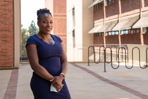
Oluwatayo (Tayo) Ikotun, Ph.D.
- Assistant Professor, Molecular and Medical Pharmacology
- Assistant Professor, Crump Institute for Molecular Imaging

Oluwatayo (Tayo) Ikotun, Ph.D., seeks to develop novel nuclear imaging tools to enhance our understanding of how the immune system responds to diseases and treatments. Her research could inform the development of precision therapeutics for patients with Type 1 diabetes, progressive fibrosis and cancers that do not respond to current immunotherapies.
Ikotun seeks to deliver off-the-shelf nuclear imaging tools that facilitate non-destructive, longitudinal and quantitative monitoring of the immune system. The effectiveness of existing and new drug products relies on the body's immune response, both at the specific tissue level and throughout the entire immune system. Thus, tools Ikotun is developing may be utilized for disease management, treatment planning, therapy monitoring and informing underlying mechanisms of action and may serve as predictive biomarkers of patient response.
Her long-term goal is to move toward multiplex in vivo imaging to facilitate global assessment of the cross-talk between distinct immune cell populations and enable comprehensive phenotypic stratification of immune subtypes. She uses positron emission tomography, or PET, and single photon emission computed tomography, or SPECT, imaging to examine how the concentration, distribution and function of immune cells change as a tumor develops and spreads in the body. Leveraging multiplex SPECT techniques that allow her to observe two to three different immune cell types simultaneously, Ikotun aims to capture a fuller picture of the various interactions taking place within the tumor microenvironment to offer a more nuanced look at a patient’s cellular response to disease and therapeutic intervention.
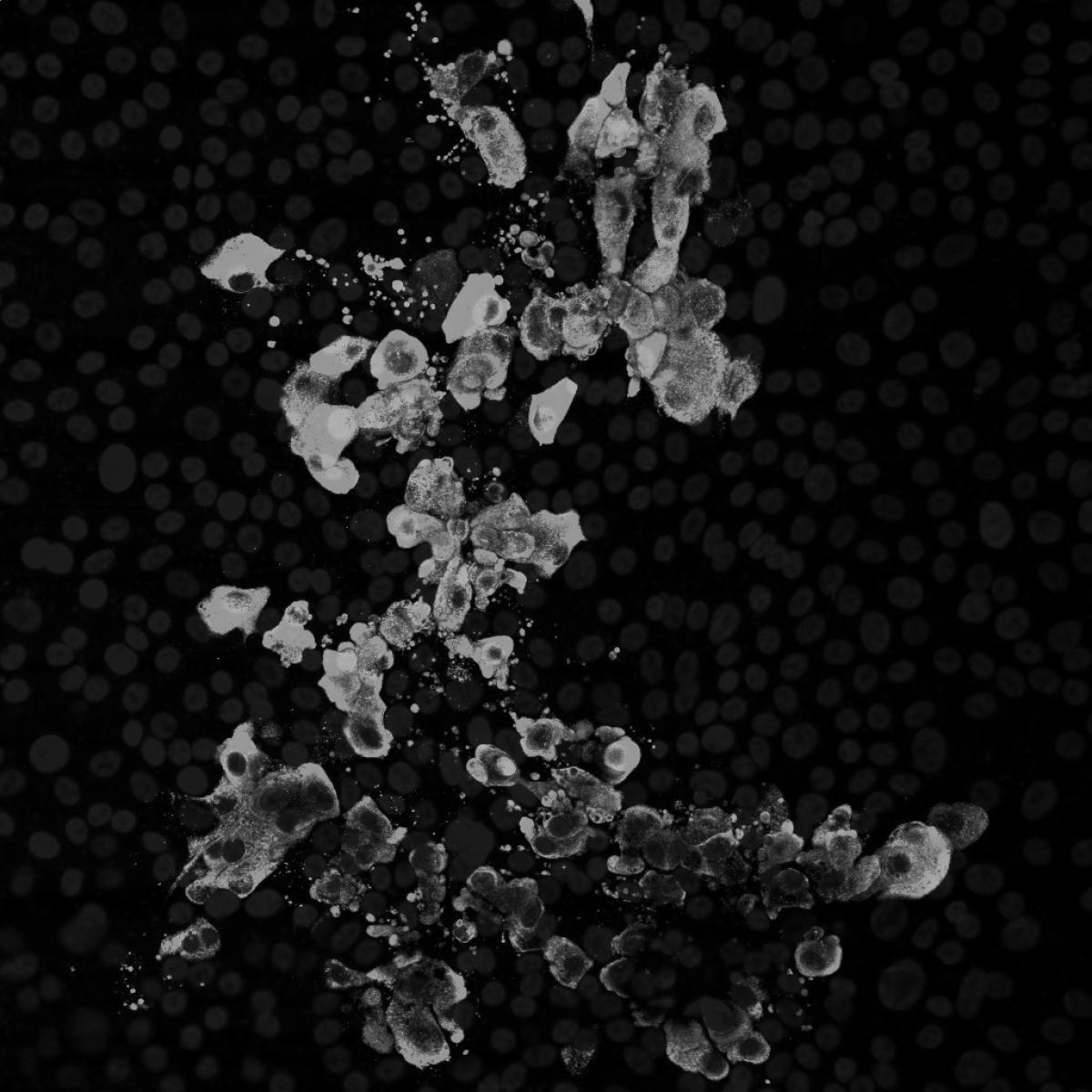
Ikotun is also developing new animal models of progressive fibrosis to support the creation of new therapies. Fibrosis can occur in nearly every part of the body and result in organ failure or death if untreated. In the past 30 years, approximately 90% of therapies that have shown promise in animal models of fibrosis have failed in human clinical trials. To create fibrosis models that more accurately reflect human disease, Ikotun implants stem cell induced fibroblast activation organoids into mice. These models can reveal how fibrosis progresses and be used to test possible treatments.
Research Projects
- Developing diagnostic imaging tools to enable in vivo A process, procedure or study performed on or in a living organism. in vivo A process, procedure or study performed on or in a living organism. visualization of the cancer immunity cycle
- Investigating the validity of a single-agent targeted radio-immunotherapy approach to overcome challenges in the effectiveness of current cancer immunotherapies
- Identify biomarkers and new therapies for progressive fibrosis Excessive scarring within an organ due to disrupted healing. It can lead to organ dysfunction and is associated with conditions like chronic kidney disease, liver cirrhosis and heart failure. fibrosis Excessive scarring within an organ due to disrupted healing. It can lead to organ dysfunction and is associated with conditions like chronic kidney disease, liver cirrhosis and heart failure. with a particular focus on interrogating fibroblast activation, macrophage dysregulation and collagen production
-
Post-doctoral Fellowship
- Radiochemistry, Molecular Imaging, Washington University School of Medicine, 2013
Degree
- Ph.D., Bioinorganic Chemistry, Syracuse University, 2009
-

