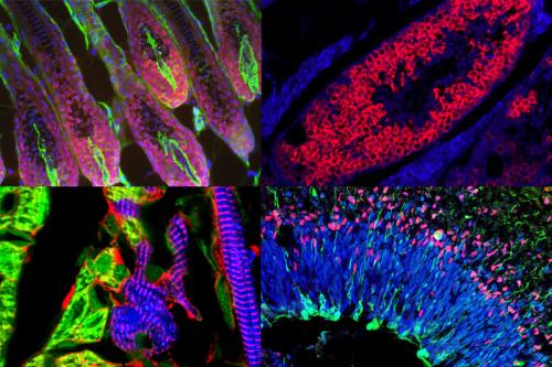
Seeking exceptional microscopy images and videos – enter the contest!
The Eli and Edythe Broad Center of Regenerative Medicine and Stem Cell Research at UCLA is announcing a light microscopy image and video contest to recognize and celebrate the amazing advances in science achieved through microscopy at UCLA.
Prizes will be awarded to submissions that exhibit exceptional structure, color, composition, informational content, and visual impact.
The contest is a collaboration of microscopy cores across campus, including: the Broad Stem Cell Research Center-Department of Molecular, Cell, and Developmental Biology Microscopy Core; the California NanoSystems Institute Advanced Light Microscopy and Spectroscopy Laboratory; and the Stein Eye Institute Microscopy Core.
Eligibility:
The contest is open to anyone at UCLA. Images or videos must have been captured using instruments located at UCLA. Videos can be any movie or digital time-lapse photography taken through a microscope at UCLA.
Prizes will be awarded for the following categories:
- Images (first, second and third place)
- Videos (first and second place)
- Zeiss prize (sponsored by Zeiss for an image generated using one of their instruments)
- Leica prize (sponsored by Leica for an image generated using one of their instruments)
- Viewers’ choice award
Prizes include: An Apple watch, iPads, Bluetooth speakers, wireless headphones, Zeiss binoculars, and $1,000 in reagents for your lab.
Submissions must include the following information to be eligible for prizes:
- Your UCLA email address
- Name(s) of the individual(s) who produced the image or video
- Name of your principal investigator (if different than individual who produced the image/video)
- Technique and Instrument used to capture the image or video
- Name of core facility used (where applicable)
- Magnification
- Title
- Description of image or video in lay-friendly language that explains: what is pictured (e.g. tissue type, cells pictured and if they are distinguished by color, process or phenomenon that is seen) and why this is being studied (what is your research aim or what is the scientific significance of the image)?
Example of a lay-friendly description: Microscopic image of a mini brain organoid, showing layered neural tissue and different groups of neural stem cells (in blue, red and magenta) giving rise to neurons (green). This mini brain organoid was created to help our lab study how the Zika virus destroys neural stem cells.
Only complete submissions will be considered.
Submit images or videos here. The submission site will be open until December 20, 2019.
Please note: By submitting to this contest, you give the UCLA Broad Stem Cell Research Center non-exclusive permission to use your submissions for communications and development purposes.