
A lesser-known SARS-CoV-2 protein may offer clues to long COVID symptoms
Key Takeaways
- UCLA researchers discovered that a lesser-known SARS-CoV-2 protein called nucleocapsid can amplify inflammatory signals in macrophages, immune cells that help detect viruses and coordinate the body’s early response.
- Using engineered macrophages and stem cell-based models, the team found that inflammatory signals from these immune cells can weaken blood-vessel barriers in the heart, illustrating one way COVID-19 can contribute to cardiac complications.
- The findings point to a potential target for future therapies designed to prevent the runaway inflammation behind severe COVID-19 and its lingering aftereffects.
Six years after the height of the COVID-19 pandemic, scientists are still uncovering surprising ways the virus can wreak havoc on the body.
A new UCLA study published in Science Advances describes a previously unrecognized way the SARS-CoV-2 nucleocapsid protein — a structural protein less familiar than the spike protein that has dominated public attention and much vaccine research — can push the immune system into dangerous overdrive.
The nucleocapsid protein's main job is to package and protect the virus's genetic material. Like several other coronavirus proteins, it's also known to suppress the body's early antiviral defenses, helping the virus get a foothold.
Scientists led by virologist Dr. Melody Li set out to see whether SARS-CoV-2's version of this immune-dampening protein worked the same way as those found in SARS and MERS, earlier coronaviruses known to cause severe disease.
“Coronaviruses are notorious for encoding proteins that antagonize the body's natural antiviral defenses,” said Li, an associate professor of microbiology, immunology and molecular genetics and member of the UCLA Broad Stem Cell Research Center. “When SARS-CoV-2 first appeared, almost nothing was known about it, so we wanted to find out whether it was using the same playbook.”
A hidden protein with an outsized effect
The project, initially funded through a COVID-19 seed grant with UCLA colleague Alexander Hoffmann, led the researchers to a less explored question: how the nucleocapsid protein behaves inside macrophages. These immune cells patrol tissues for signs of infection and release chemical signals called cytokines and chemokines to rally the body’s defenses.
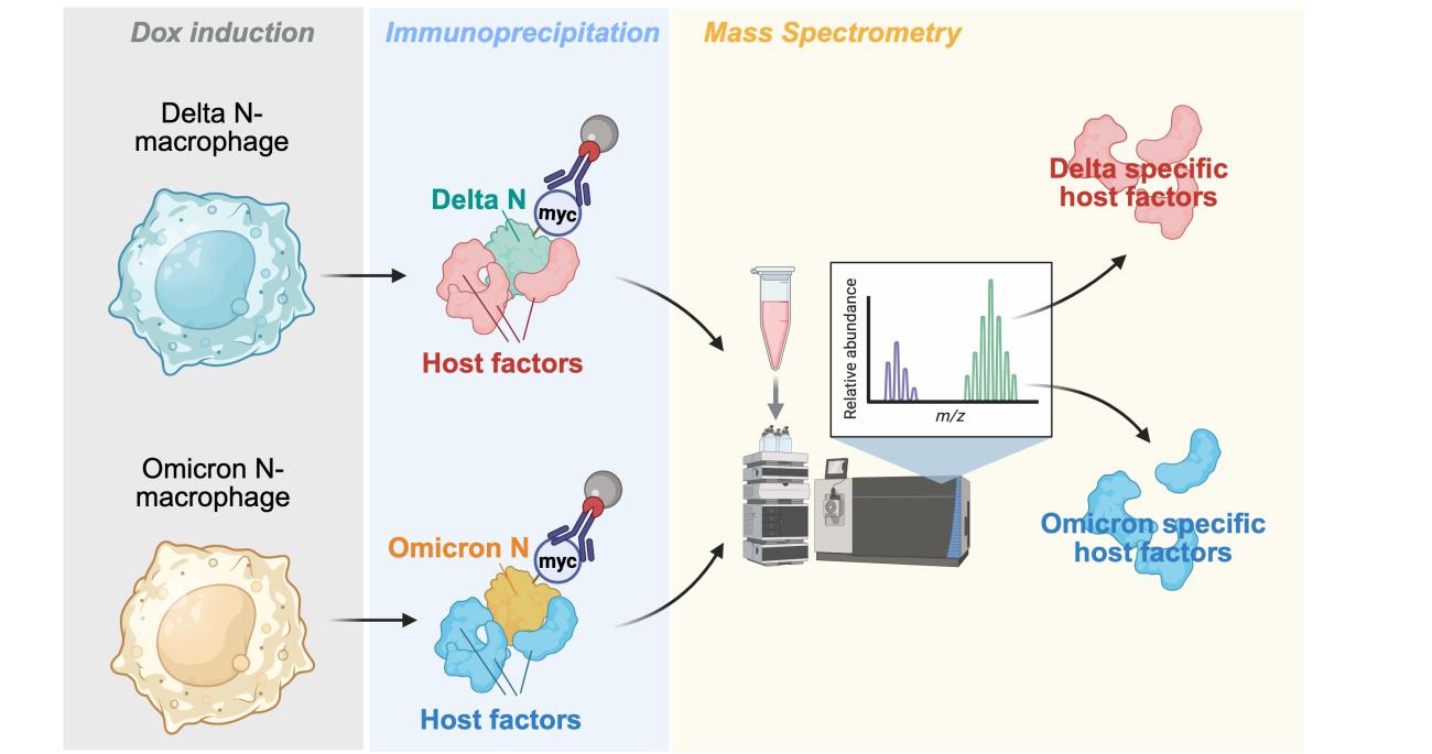
What they found upended their original hypothesis. The nucleocapsid protein appeared to be a “double-edged sword,” Li said. While it still suppressed signals that trigger an early antiviral response, it also amplified inflammatory pathways in macrophages, which can fuel tissue-damaging immune responses.
“We set out looking for a protein that suppresses the immune response, and we found the opposite,” said Dr. Zhenlan Yao, co-first author of the study and a former postdoctoral researcher in Li’s lab, who will soon begin a research assistant professorship at Hong Kong University. “It was surprising, but it lines up with what we already know about COVID-19: The virus dampens the immune response early on, then overactivates it later — and that’s when a lot of the tissue damage happens.”
The researchers examined nucleocapsid proteins from several SARS-CoV-2 variants, as well as from SARS-CoV-1 and MERS-CoV, and found the pro-inflammatory effect was conserved across pathogenic coronaviruses — with the Delta variant’s version proving the most inflammatory by far.
“It’s a bit like a thief trying to slip past a bank’s security system, but instead of staying quiet, it trips the alarm,” Li said. “We don’t think these viruses intend to do this — a virus’s whole goal is to spread, not to make its host severely sick. But this looks like an unintended side effect that in the case of COVID, it ends up fueling the disease.”
In the body, that immune “alarm” does not stay confined to macrophages. When these cells become overactivated, they release inflammatory signals that can affect nearby tissues, including the cells that line blood vessels.
Cracks in the body's protective barriers
To investigate whether those effects could help explain COVID-19 complications involving the brain and heart, the team turned to two human cell-based models: a stem cell-derived model of the blood-brain barrier and a model of the coronary artery lining.
These barriers are made of endothelial cells, which line blood vessels and help control what passes from the bloodstream into surrounding tissues. In the brain, this barrier is especially tight, helping protect delicate neural tissue from pathogens, toxins and other harmful substances.
When the researchers exposed both models to fluid containing signals from macrophages producing the Delta variant’s nucleocapsid protein, the heart barrier broke down significantly — a phenomenon known as vascular leakage.
Because the heart depends on tight, selective blood vessel linings to function normally, the finding points to a possible mechanism that could help the cardiac injury seen in severe cases of COVID-19.
Rethinking how severe COVID-19 is treated
The findings also suggest a path toward more targeted COVID-19 treatments.
Severe cases can be treated with broad anti-inflammatory drugs like corticosteroids, which dampen harmful inflammation but do not specifically target the viral mechanisms that may be driving it. A therapy or vaccine that targets the nucleocapsid protein, Li said, could potentially rein in the hyperinflammation more precisely — and, in doing so, help protect the blood vessel barriers that support brain and heart health.
And because macrophages play a similar double-edged role in many infections beyond COVID-19, she said, the same mechanism could turn out to matter well beyond this one virus.
“It’s critical to keep studying COVID-19 so that we can constantly improve patient care — not everyone responds well to vaccines, and people who are immunocompromised often have limited treatment options,” said Pablo Alvarez, co-first author of the study and a former graduate student in Li’s lab. “These studies can also help us prepare for future coronavirus outbreaks.”
Additional UCLA authors include Carolina Chavez, Yennifer Delgado, Prashant Kaushal, David Austin, Qian Li, Yanying Yu, Anne Zaiss, Vaithilingaraja Arumugaswami, Jeffrey Hsu, Robert Damoiseaux, Mehdi Bouhaddou and Alexander Hoffmann. Qiang Din of the Tsinghua University also contributed to this study.
Notes
This work was supported by the National Institute of Allergy and Infectious Diseases, the W.M. Keck Foundation and the American Heart Association. Additional awards were provided by the David Geffen School of Medicine at UCLA; the UCLA Health Jonsson Comprehensive Cancer Center and UCLA Broad Stem Cell Research Center Ablon Scholars Program; and the UCLA Department of Microbiology, Immunology and Molecular Genetics.
Contact
Meet the Researchers

- Associate Professor, Microbiology, Immunology and Molecular Genetics
How viruses manipulate cells
This study fits into a broader pattern emerging from Li’s lab.
In a 2024 study, Yao, co-first author of the current study, showed that structural proteins from chikungunya virus — a mosquito-borne pathogen unrelated to coronaviruses — can also influence how the virus behaves inside immune cells. In that study, the proteins helped chikungunya virus infect macrophages more efficiently by interacting with host immune proteins.
Structural proteins like these were traditionally thought to matter mainly for helping viruses enter and exit cells, not for manipulating the immune system from within.
“Both of these projects point to a role for viral structural proteins in interacting with and even modulating the innate immune response — something we’re only beginning to appreciate,” Li said. “It’s part of a bigger theme we’re seeing across very different viruses, and I expect we’ll learn a lot more about it in the coming years.”