
Announcing the winners of the Broad Stem Cell Research Center’s 2023 microscopy image and video contest
The Eli and Edythe Broad Center of Regenerative MedicineA field focused on developing and applying new therapies and techniques to repair, replace or regenerate tissues and organs and restore function that has been lost due to aging, disease, injury or genetic defects.Regenerative MedicineA field focused on developing and applying new therapies and techniques to repair, replace or regenerate tissues and organs and restore function that has been lost due to aging, disease, injury or genetic defects. and Stem Cell Research at UCLA is pleased to announce the winners of its 2023 light microscopy image and video contest.
For the second time, the center is honoring the remarkable work achieved through microscopy by researchers from across UCLA. The level of skill and creativity displayed by all contest participants was inspiring. After careful evaluation by a panel of scientists and microscopy experts, the six winning entries were selected for exhibiting a combination of technical expertise, visual impact and scientific relevance.
Here are this year’s winners:
First place image
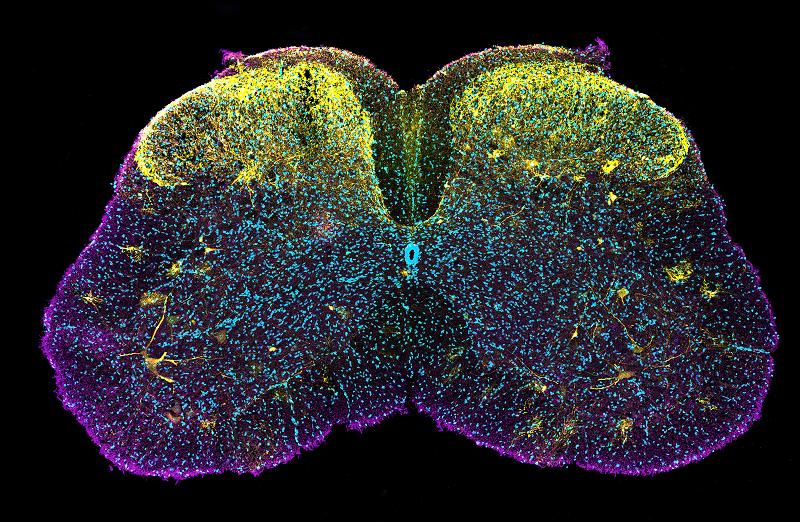
Title: Spinal starry night
Instrument: Zeiss LSM800
Magnification: 10x
Creator: Soizic Riche, Ph.D., a postdoctoral scholar in the lab of Samantha Butler, Ph.D.
This image shows the expression of the protein cofilin (yellow) in specific neuronal subpopulation of a juvenile mouse spinal cord (magenta and cyan). Cofilin plays a key role in the process by which developing nerves extend thread-like projections called axons over long distances to form neural networks throughout the body. A better understanding of this process could lead to the creation of treatments that reduce the time it takes for people to recover from peripheral nerve injuries.
Second place image
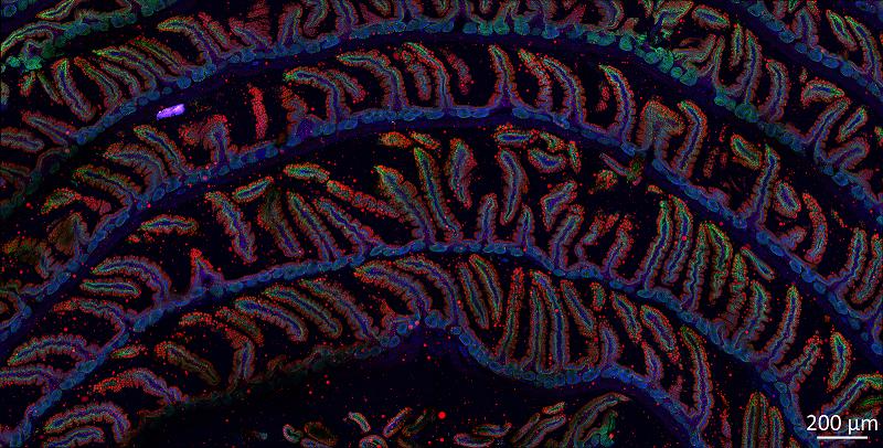
Title: Intestine rainbow
Instrument: Zeiss LSM 900
Magnification: 40x
Creators: Yajing Gao, Ph.D., a postdoctoral scholar in the lab of Peter Tontonoz, M.D., Ph.D. and Julia Mack, Ph.D.
Microscopic image of the side view of a "swiss roll" of a mouse small intestine, showing accumulation of lipids in droplet structures (red) within the cells (blue) of the intestine lining (green) after the mouse had gorged on a high fat meal. This image was created to aid in the study of how metabolic pathways affect fat absorption in the intestine. Understanding this process is important for developing interventions for obesity and treating metabolic diseases such as hyperlipidemia.
People's choice image
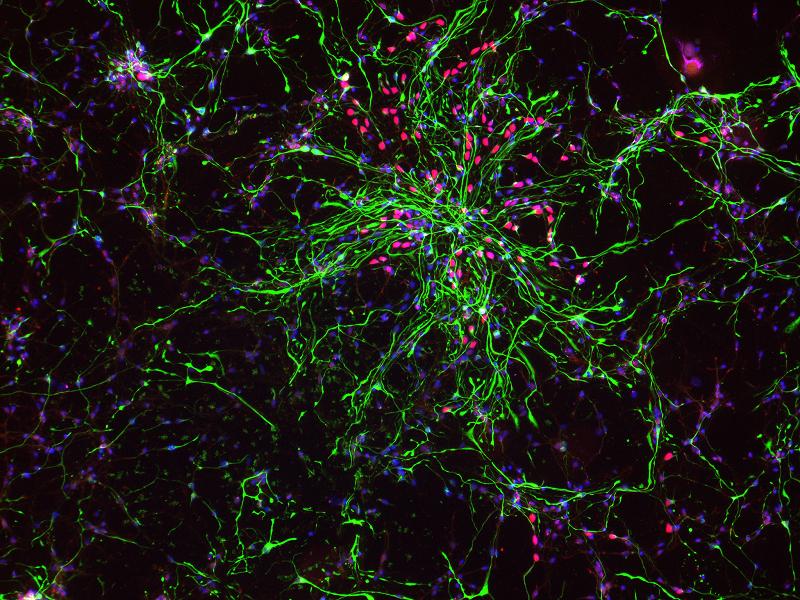
Title: A glimpse inside the developing brain
Instrument: EVOS M5000 by Invitrogen ThermoFisher Scientific
Magnification: 10x
Creator: Jessenya Mil, a Broad Stem Cell Research Center trainee and graduate student in the lab of Aparna Bhaduri, Ph.D.
The Bhaduri lab aims to understand the building blocks that contribute to the development of the human cortex, the outermost surface of the brain. There are several types of cells that arise during this development, such as astrocytes (green) and intermediate progenitor cellsDescendants of stem cells that can further differentiate to produce one or more specialized cell types. They are more limited than pluripotent stem cells in that they cannot self-renew indefinitely and can only produce a limited range of specific cell types. For example, neural progenitor cells can only produce neurons.progenitor cellsDescendants of stem cells that can further differentiate to produce one or more specialized cell types. They are more limited than pluripotent stem cells in that they cannot self-renew indefinitely and can only produce a limited range of specific cell types. For example, neural progenitor cells can only produce neurons. (red). Understanding the cell types, and therefore the building blocks, of the developing human cortex allows the lab to identify origins of neurodevelopmental disorders and highlight therapeutic targets to avoid the onset of these disorders.
Best undergraduate submission
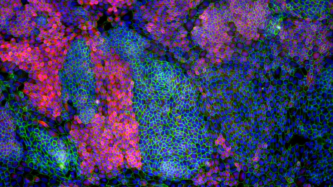
Title: Signature protein expression of mature retinal pigment epithelium cells
Instrument: Olympus BX51
Magnification: 20x
Creators: Caitlin Chen, an undergraduate student, and Shahab Younesi, a core scientist, at the UCLA Broad Stem Cell Research Center’s Translational Cell Therapy Lab led by Anupama Dimashkie
This image captures a retinal pigment epithelium, or RPE, derived from an induced-pluripotent stem cell, or iPSC, line. Image shows cell borders (green), mature RPE-specific marker (red), and cell nuclei (blue) in iPSC-RPE cells in order to confirm the identity of iPSC-derived RPE cells. The RPE is a single cell layer in the retina that is essential for photopic vision, so progressive RPE degeneration severely compromises visual function. Stem cell-derived RPE cells are considered a promising treatment for RPE atrophy. The research goal of the Translational Cell Therapy Lab is to characterize iPSC-RPE cells for safety and functionality before clinical application into a patient’s subretinal space for vision restoration.
First place video
Title: Microglia unleashed: dynamic response to a laser injury
Instrument: Leica Stellaris 8 DIVE
Magnification: 25x
Creator: Fanny Etienne, Ph.D., a postdoctoral researcher in the lab of Lindsay De Biase, Ph.D.
This video of acute mouse brain slices depicts microglia cells, the immune cells of the central nervous system, responding to a laser injury (white circle at center) over 30 minutes after the lesion. Microglia are constantly extending and retracting thread-like projections called processes that allow them to survey their environment and respond to any injury or infection in the brain. The De Biase lab uses imaging of this kind to study how aging can disrupt microglia’s surveillance and response to injury.
Second place video
Title: Playing with your food
Instrument: Nikon Ti2 Spinning Disk Confocal Microscope
Magnification: 60x
Creator: Gil Torten, a graduate student in the lab of David Williams, Ph.D.
Retinal pigment epithelium cells, which line the inner surface of the back of the eye, ingest outer segments of photoreceptor cells, the rods and cones in the retina that respond to light, to maintain visual health. When this ingestion process stops, photoreceptors die, resulting in blindness. In this 10-minute clip that has been condensed into three seconds, two mouse retinal pigment epithelium cells (red) pass an outer segment (pink) back and forth, ostensibly playing with their food.
The center would like to thank all who submitted images and videos as well as the contest judges, including Mark Terasaki, Ph.D., who also supported a portion of this contest through generous philanthropy dedicated in memory of his father, Dr. Paul Terasaki.
To see all contest submissions, visit the center's Flickr. The 12 images that received honorable mentions are listed first in the album.